

Accurate Radiological Diagnosis of Ovarian Torsion at Kailash Hospital and Heart Institute, Noida Sector - 27
A 21 yr old, unmarried female presented in emergency department with sudden onset, severe abdominal pain. She had no gastrointestinal or genitourinary complaints. She had normal regular menstrual cycles. There was no significant past medical or surgical history. She had not previous ultrasound done.
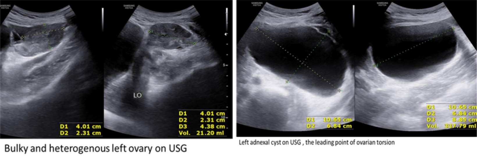
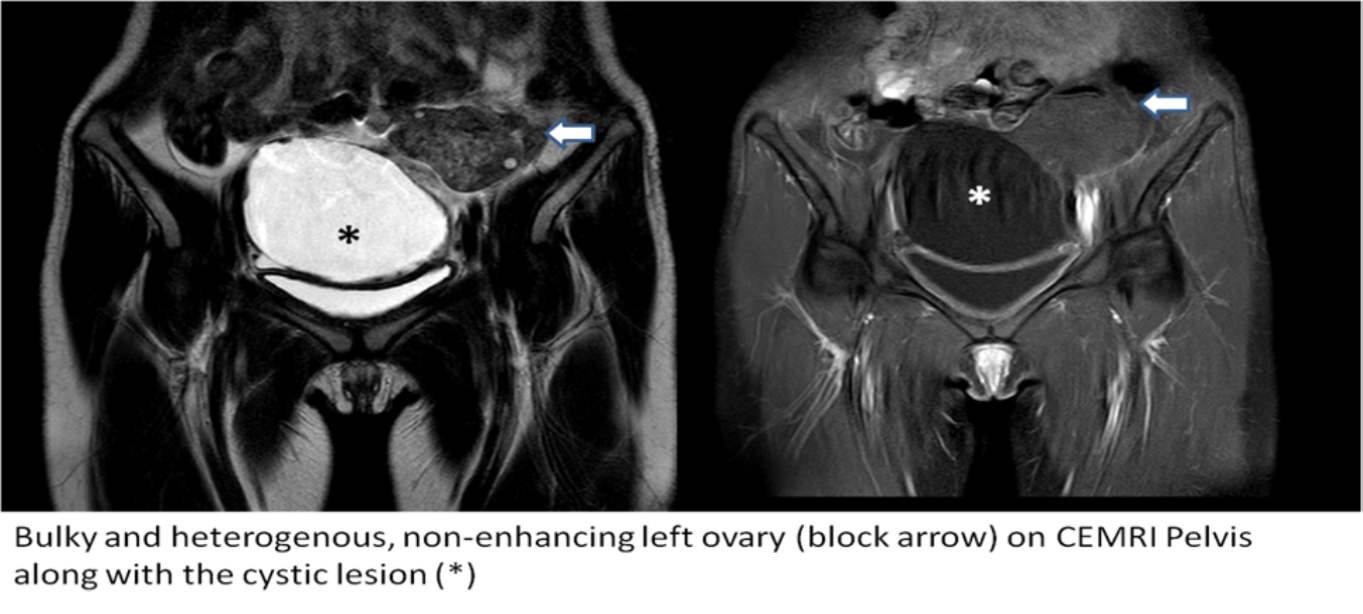
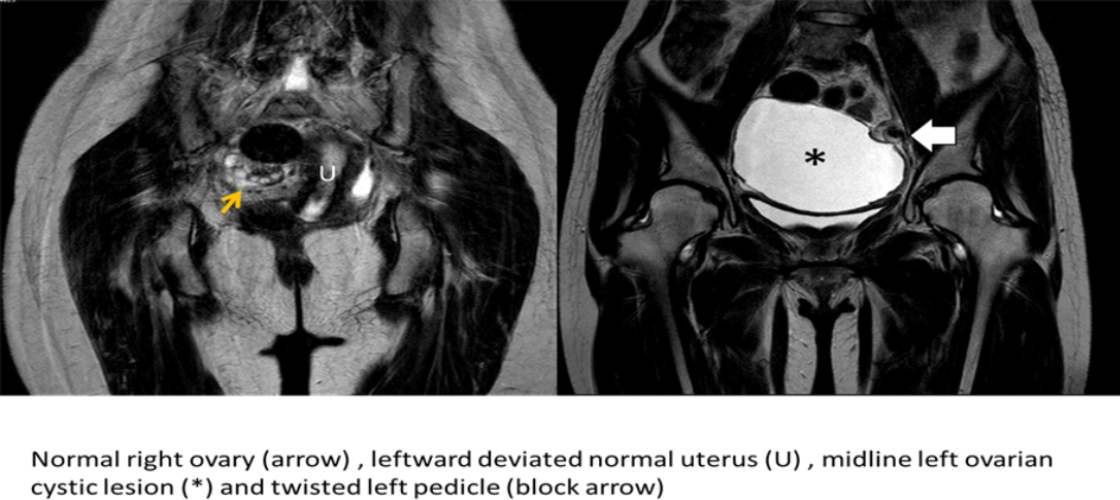
She underwent a transabdominal ultrasound evaluation in Radiology department, headed by Dr. Kanchan Varma. The scan was performed by Dr. Shivani Gupta (Consultant Radiologist) which revealed a large exophytic left ovarian simple cyst of size 10 x 9 cm with no obvious septations or solid component. The cyst was located in midline abutting the uterus. The left ovarian parenchyma adjacent to the cyst was grossly bulky and heterogenous with tiny peripheral follicles. Color Doppler did not reveal any vascularity within left ovary or cyst walls. The uterus and right ovary were normal in appearance. Based on the clinical profile and ultrasound findings probability of left ovarian torsion was raised. Patient was further advised CEMRI Pelvis for confirmation as TVS evaluation was not possible. CEMRI revealed thickened twisted left ovarian pedicle with enlarged, heterogenous, non enhancing left ovarian parenchyma suggesting non viable left ovary in addition to the large ovarian cyst seen previously on ultrasound examination. The uterus was seen deviated towards left side. Based on these findings, the diagnosis of left ovarian torsion was established.
The patient underwent emergency surgery by Dr.(Col) Sunil Chawla (Obstetrician and Gynecologist). The per operative findings were consistent with the imaging findings which showed a large left exophytic ovarian cyst twisted twice on its own pedicle along with non viable ovary and hence oophorectomy was performed. The patient was stable at discharge the next day.
Ovarian / adnexal cyst torsion is a gynecologic surgical emergency. It occurs when the ovary rotates around its supporting ligaments, twisting and compressing the accompanying blood vessels and lymphatics. The diagnosis can be missed which can lead to delay in treatment and complications.
Verified by :

Dr. Kanchan Varma
Dr. Kanchan Varma is the Head of the Department (HOD) of Radiology at Kailash Hospital, Noida. With an MD in Radio Diagnosis, she specializes in advanced imaging techniques and diagnostics. Her expertise in radiology plays a crucial role in accurate disease detection and treatment planning. Dr. Varma is dedicated to providing precise and high-quality diagnostic services, ensuring optimal patient care through cutting-edge radiological advancements.